
Rapid Calcification of Myocardium as Sequela from Severe Sepsis
Article Information
- Received
- Accepted
- Published
Academic Editor: Anil TOMBAK, Mersin University Faculty of Medicine
Checked for plagiarism: Yes
Review by: Single-blind
Copyright © 2017 Andreas S. Kunz, et al
 This is an open-access article distributed under the terms of the Creative Commons Attribution License, which permits unrestricted use, distribution, and reproduction in any medium, provided the original author and source are credited.
Corresponding author: Andreas S. Kunz, University Hospital Wurzburg, Department of Diagnostic and interventional Radiology —
Competing Interests
The authors have declared that no competing interests exist.
Funding
No specific funding statement was provided by the authors.
Data Availability
No data-availability statement was provided by the authors.
Citation:
The Case
The reported case shall highlight severe sepsis as a possible cause of myocardial calcification, as well as the highly dynamic development thereof within a time span of merely 10 weeks. A 60-year-old male patient had been admitted to hospital for palliative therapy of progressive multiple myeloma, which lately had transformed into plasma cell leukemia. After commencing his third therapy cycle with Elotuzumab, the patient suffered from a pneumogenic sepsis due to staphylococcus infection that required mechanical ventilation for 7 days. Antimicrobial therapy followed antimicrobial susceptibility testing and included Tazobactam, Piperacillin, and Fosfomycin. During his hospital stay, computed tomography images of the chest were acquired initially, i.e. at onset of pneumogenic sepsis (Figure 1.: left panel), as well as after 10 weeks (Figure 1: right panel) to follow-up pneumonia consolidations in both upper lung lobes. Surprisingly, non-contrast enhanced follow-up CT images revealed newly developed calcifications within the outer myocardial layers of the left ventricle.
Figure 1. Non-contrast enhanced CT scans of the chest initially during onset of pneumogenic sepsis (right) and after 10 weeks (left) show rapid development of non-preexisting, extensive myocardial calcifications.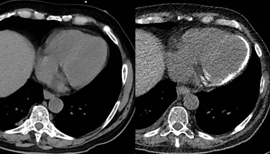
Download figure
Only very few reports exist describing myocardial calcification as a sequela from severe sepsis. Explanations attribute alterations of myocardial microcirculation to cause subsequent tissue necrosis during septic shock and capillary leak and relative capillary stasis.1 Resulting interstitial and intracellular edema, and consecutive mitochondrial destruction and cellular necrosis.2 In other cases, myocardial calcifications have been described as a complication resulting from myocarditis.3
In general, myocardial calcifications are associated with myocyte necrosis due to severe infection or inflammation, and can lead to restrictive cardiomyopathy. Differential diagnoses include calcifications of the inner myocardial layers, which can be detected after myocardial infarction, and pericardial calcifications that indicate constrictive pericarditis.
References
Cited by (5)
This article has been cited by 5 scholarly works according to:
Citing Articles:
Radiology: Cardiothoracic Imaging (2021) Crossref OpenAlex
Joy Li, Lydia Chelala, Rydhwana Hossain, J. Jeudy, C. White - Radiology: Cardiothoracic Imaging (2021) Semantic Scholar
Journal of Forensic and Legal Medicine (2020) Crossref OpenAlex
Simone Cappelletti, D. Piacentino, C. Ciallella - Journal of Forensic and Legal Medicine (2020) Semantic Scholar
BMJ Case Reports (2019) Crossref OpenAlex Semantic Scholar